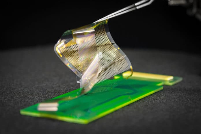
A team of engineers and neurosurgeons from the University of California San Diego Jacobs School of Engineering has developed a new array of brain sensors that can record electrical signals directly from the surface of the human brain in record-breaking detail. High-resolution recordings of electrical signals from the surface of the brain could improve neurosurgeons’ ability to remove brain tumors and treat epilepsy and could open up new possibilities for medium- and longer-term brain-computer interfaces.
The new device is a type of electrocorticography (ECoG) sensor, which is already in common use as a tool by neurosurgeons performing procedures to remove brain tumors and treat epilepsy in people who do not respond to drugs or other treatments. The ECoG grids most commonly used in surgeries today typically have between 16 and 64 sensors, although research-grade grids with 256 sensors can be custom-made.
Thanks to some important engineering advances, the UC San Diego team was able to produce ECoG grids with either 1,024 or 2,048 sensors. If approved for clinical use, these thin, pliable grids of ECoG sensors would offer neurosurgeons brain-signal information directly from the surface of the brain’s cortex in 100 times higher resolution than what is available today.
The team was able to produce grids with a far greater density by using nanoscale platinum rods, which offer more sensing surface area than flat platinum sensors used today. The new platinum nano-rod brain sensor grids are ten micrometers thick, approximately one-tenth the size of a human hair, and 100 times thinner than the one millimeter thick and clinically approved ECoG grids. This provides the new grids with 100 sensors per unit area compared to 1 sensor per unit area for clinically used grids, offering 100 times better spatial resolution in interpreting brain signals. The nano-rods are embedded in a transparent, soft, and flexible biocompatible material called parylene, which is in direct contact with the surface of the brain.
In demonstrations, the team’s three-centimeter-by-three-centimeter grid with 1,024-sensors recorded signals directly from the brain tissue of 19 people who agreed to participate in this project during the “downtime” of their already scheduled brain surgeries related to either cancer or epilepsy. In particular, the team developed functional maps in four different people of a boundary in the brain called the central sulcus during motor tasks. They also mapped the cortical column of a rat brain for the first time without the use of a needle and electrical stimulation.
The team is working on a range of initiatives in parallel to advance these grids so that they are eligible for review for approval for short-, medium- and longer-term use.
