
A potentially bone-breaking mishap usually requires an X-ray examination of the injured limb and a trip to the nearest hospital or emergency clinic. However, soon, imaging services can be brought closer to the patient.
Researchers at Finland’s University of Oulu are currently developing a compact X-ray imaging device that even a patient could use himself if necessary. According to a university release, there is now a prototype of the automated X-ray machine that will soon be ready for patient testing.
The current large X-ray units require lead protection for the entire imaging room, which is expensive and takes up space. But, the new portable X-ray machine measures only approximately 50x50x130 cm, thanks to the built-in radiation protection.
It is easy to use and can, in principle, be stored and used anywhere it might be necessary, such as a municipal center, emergency clinic, or a ski resort – which is often a long way to the nearest hospital or on-call health center to check for possible fractures caused by a fall on a slope. This means that the device can be used not only to improve the accessibility of health services but also to achieve significant cost savings in health care and specialized medical care.
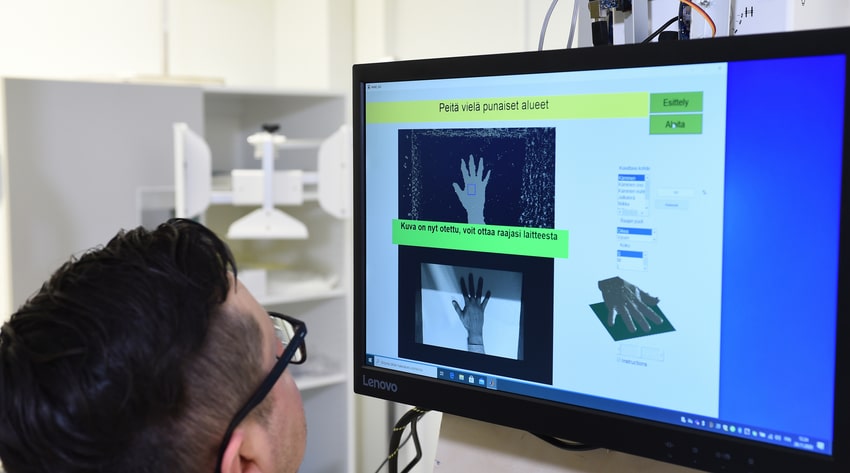
With this machine, the self-diagnosis process is guided by the computer at all times. At this stage of development, imaging voltage and guidance modeling of the self-guiding X-ray have been scaled for X-raying bones in the ankle and palm. The actual innovation of the device is automated patient control, which guides the patient to set the injured limb in the correct position for imaging.
The X-ray imaging device is said to improve the accessibility of health services and bring cost savings. Additionally, it can be used to lighten the workload of trained X-ray personnel in health centers and university hospitals and to allocate these resources for more demanding imaging tasks.